Medical Imaging Segmentation
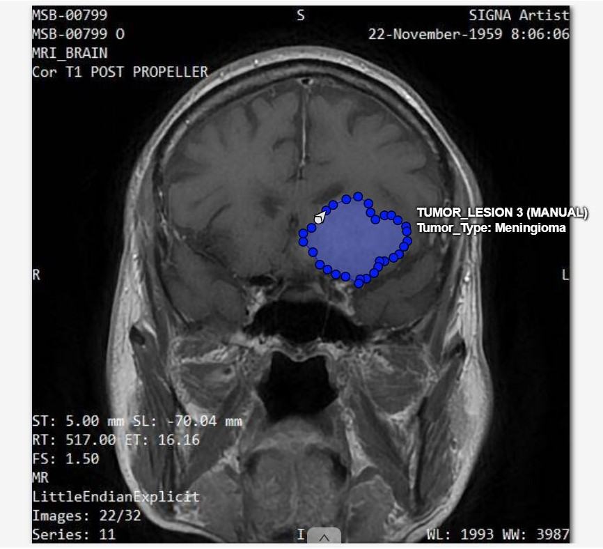
Image 1 — Coronal T1 POST MRI scan. Blue polygon: manually annotated Meningioma tumour lesion (TUMOR_LESION 3). The annotator isolated the tumour boundary using the polygon tool, excluding peripheral vasogenic edema, per our internal annotation guideline.
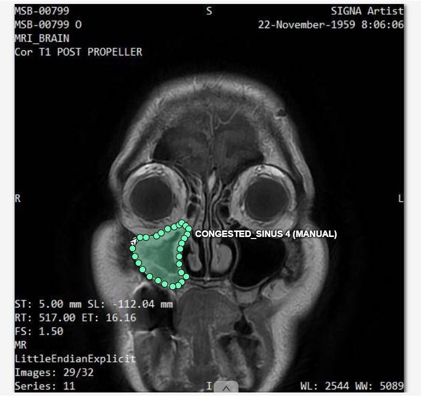
Image 2 — Same patient, inferior axial slice. Teal polygon: manually annotated congested sinus region (CONGESTED_SINUS 4). Both scans are from the same multi-image MRI series, demonstrating multi-slice annotation consistency.
Healthcare AI
Medical Imaging Segmentation
Pixel-level segmentation of radiology scans (CT, MRI, X-ray) demonstrating our polygon-based annotation and multi-slice QA workflow on publicly available imaging data.
What You're Seeing
Two slices from the same coronal MRI brain series (MSB-00799). Image 1 shows a Meningioma tumour annotated with a blue filled polygon at the mid-brain level. Image 2 shows an inferior slice where a congested sinus region is annotated in teal. Both annotations were drawn manually using the polygon tool and cross-referenced against adjacent slices to ensure volumetric accuracy.
Annotation Type
Pixel-level polygon segmentation · Tumour boundary isolation · Multi-slice volumetric consistency checks
Annotation Rule
Annotators isolated the tumour core excluding peripheral vasogenic edema, per our internal annotation guideline. Each boundary was verified against adjacent slices for 3D volumetric continuity.
QA Standard
Dice Similarity Coefficient tracked per case — any score falling below the required threshold escalated to a senior annotator for re-review.